Mhuji Kilonzo,
Patrick A Ndakidemi,
Musa Chacha ![]()
For correspondence:- Musa Chacha Email: musa.chacha@nm-aist.ac.tz Tel:+255753458177
Received: 14 June 2016 Accepted: 16 September 2016 Published: 31 October 2016
Citation: Kilonzo M, Ndakidemi PA, Chacha M. In vitro antifungal and cytotoxicity activities of selected Tanzanian medicinal plants. Trop J Pharm Res 2016; 15(10):2121-2130 doi: 10.4314/tjpr.v15i10.10
© 2016 The authors.
This is an Open Access article that uses a funding model which does not charge readers or their institutions for access and distributed under the terms of the Creative Commons Attribution License (http://creativecommons.org/licenses/by/4.0) and the Budapest Open Access Initiative (http://www.budapestopenaccessinitiative.org/read), which permit unrestricted use, distribution, and reproduction in any medium, provided the original work is properly credited..
Purpose: To evaluate the antifungal and cytotoxic activities of four medicinal plants from Tanzania, namely, Mystroxylon aethiopicum, Lonchocarpus capassa, Albizia anthelmentica and Myrica salicifolia.
Methods: The plant materials were subjected to extraction using dichloromethane, ethyl acetate and distilled water. The minimum inhibition concentration (MIC) of the extracts against Candida albicans and Cryptococcus neoformans was determined by microdilution method. The lowest concentration which showed no fungal growth was considered as MIC. The cytotoxic effect of the extracts was determined using brine shrimp toxicity assay.
Results: Lonchocarpus capassa leaf extracts exhibited antifungal activity against test fungal strains with MIC range of 0.78 – 3.13 mg/mL with Lonchocarpus capassa aqueous leaf extract (LCAL) inhibiting C. albicans and C. neoformans at MIC value of 0.78 mg/mL. Cytotoxicity assay revealed that LCAL extract which displayed good antifungal activity, was cytotoxic against brine shrimp larvae with half-maximal lethal concentration (LC50) value of 17.86 µg/mL. Interestingly, 33 % of plant extracts exhibited high cytotoxicity with LC50 values below that of the standard anticancer drug, cyclophosphamide (16.57 µg/mL).
Conclusion: The results obtained suggest that LCAL needs to be further investigated for its phytochemical composition to unravel its antifungal secondary metabolites. Furthermore, some of the plant extracts are potential anticancer agents.
Introduction
The importance of medicinal plants in solving the healthcare problems of the world is gaining attention [1]. The World Health Organization (WHO) estimated that 80 % of the world's population has been using medicinal plants for many years as a primary healthcare [2]. Some of these medicinal plants involve the use of crude plant extracts in the form of infusion, decoction or tincture which may contain some molecules, often with indefinite biological effects [3]. Medicinal plants therefore, have been proved to be a good source of antimicrobial agents exemplified by a number of lead compounds that are currently at different stages of clinic evaluation [4].
Fungal diseases reported to be the main causes of morbidity and mortality worldwide [5]. Human infections particularly those involving skin, constitute a serious problem especially in tropical and subtropical developing countries [6]. In humans, fungal infections range from superficial to deeply invasive or disseminated [7]. According to Hamza et al [8], fungal infections particularly those caused by Candida albicans and Cryptococcus neofromans are the most challenging infections facing immune compromised patients such as HIV/AIDS patients.
Drugs currently available to treat fungal infections have serious limitations such as development of fungal resistance and toxic side effects [9]. Despite these limitations, screening for alternative means of treating fungal infections is desirable. Use of medicinal plants can be a good approach for counteracting some limitations facing conventional drugs [10]. However, most of the available information regarding the medicinal potential of these plants is not provided with scientific data. This study therefore reports the antifungal and cytotoxicity activity of M. aethiopicum, L. capassa, A. anthelmentica and M. salicifolia growing in Tanzania.
Methods
Acquisition of materials
Dichloromethane was purchased from Avantor Perfomance Materials Limited, Gujarat, India. Dimethyl sulphoxide (DMSO) and ethyl acetate were bought from RFCL Limited, Haryana, India. Sabouraud dextrose agar and Saboraud dextrose broth were supplied by HIMEDIA Laboratories Pvt. Limited, Mumbai, India. Candida albicans (ATCC 90028) and Cryptococcus neoformans (clinical isolate) were obtained from the department of Microbiology, Muhimbili University of Health and Allied Sciences (MUHAS). Standard fluconazole and iodonitrotetrazolium chloride were supplied by Lincoln Pharmaceuticals LTD, Khatraj, India and SIGMA® (Sigma- Aldrich®, St. Louis, USA) respectively. Brine Shrimps eggs were obtained from the Aquaculture innovations (Grahamstown 6140, South Africa) and sea salt was prepared locally by evaporating water collected from the Indian Ocean, along the Dar es Salaam coast, Tanzania.
Preparation of plant extracts and extraction
The plant materials were collected from different parts of Arusha region. Leaves, stems and roots of L. capassa and A. anthelmentica were collected from Esilalei village while the same plant parts of M. aethiopicum and M. salicifolia were collected from Imbibya and Engalaoni villages respectively. Plant species were identified by Mr. Gabriel Laizer, a senior botanist from Tropical Pesticide Research Institute (TPRI) and voucher specimens coded MA-01, LC-02, AA-03 and MS-04 for M. aethiopicum, L. capassa, A. anthelmentica and M. salicifolia, respectively, are kept at Nelson Mandela African Institution of Science and Technology (NM-AIST), Arusha, Tanzania. The plant materials were air-dried and pulverized into fine particles using electric blender. For non-polar and medium polar extraction, pulverized materials (250 g of leaves, stem and root barks) were sequentially successively macerated in dichloromethane and ethyl acetate for 48 h. The respective extracts were filtered through Whatman No. 1 filter paper on a plug of glass wool in a glass column and solvents were evaporated through the vacuum using a rotary evaporator. For polar extraction, the same pulverized materials (250 g of leaves, stem and root barks) were added to a 1 L of distilled water at 70 °C and allowed to cool until reaching 40 °C in a water bath. The extracts were sieved and centrifuged at 5000 rpm for 10 min. The supernatant was collected and filtered using Whatman No. 1 filter paper and dried by freezing to eliminate water by sublimation. All extracts were stored in a deep freezer at -20 °C for further activities.
Determination of antifungal activity
Minimum inhibitory concentrations (MICs) were determined by microdilution method using 96-well plates according to procedure reported by [11]. The plates were first preloaded with 50 μL of Saboraud’s dextrose broth media in each well followed by addition of 50 μL of 100 mg/mL extract (prepared in DMSO) into the first wells of each row so as to make a total volume of 100 μL in each of the first row wells. The contents were thoroughly mixed and 50 μL of the same were drawn from each of the first row wells and put into the next row wells. The process was repeated down the columns to the last wells at the bottom from which 50 μL were discarded. Thereafter, 50 μL of the selected fungal suspension (0.5 Mac Farhland standard turbidity) were added to each well thus making a final volume of 100 μL per well. Fluconazole was used in two rows of each plate to serve as standard positive control drugs against the test fungal strains while DMSO was used as negative control. Likewise, Saboraud’s dextrose broth was used to monitor fungal growth respectively. The plates were then incubated at 37 °C for 24 h. MICs for each extract were determined by adding 20 μL of 0.02 % p-iodonitrotetrazolium (INT) chloride dye in each well followed by incubation at 32 °C for 1 h. Fungal growth was indicated by change of colour to pink. The lowest concentration which showed no fungal growth was considered as MIC.
Brine shrimp lethality test
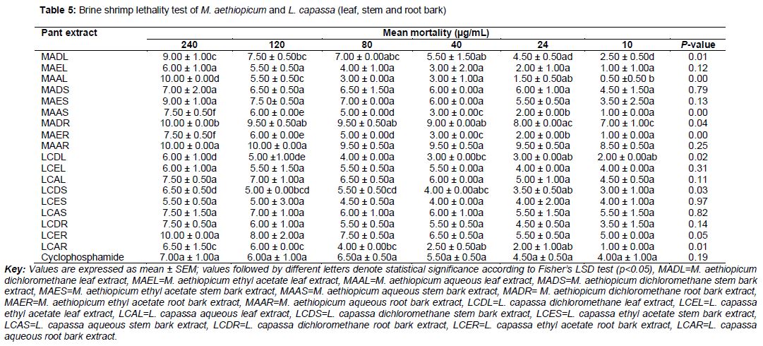
Brine shrimp (Artemia salina) larvae were used as indicator animals for preliminary cytotoxicity assay of the extracts as reported by [12]. Artificial sea water was prepared by dissolving sea salt (3.8 g) in 1 L distilled water. The salt solution was poured into a glass container and the shrimp eggs were spread and a lamp was illuminated from one side in order to attract hatched shrimps. The hatched shrimps (mature nauplii) were collected after 36 and 48 h of hatching. Stock solution of each extract was prepared by dissolving 40 mg/mL in DMSO. Different levels of concentrations (240, 120, 80, 40, 24 and 8 µg/mL) were prepared by drawing different volumes from the stock solutions and then added in a 10 mL universal bottle containing 10 brine shrimps larvae. The volume was then adjusted to 5 mL with artificial sea water prepared by dissolving 3.8 g of sea salt in 1 L of distilled water. Each level of concentration was tested in duplicate. Cyclophosphamide was used as standard positive control drug whereas DMSO and artificial sea water as negative control. The number of surviving larvae was determined after 24 h and the percentage mortality was determined by comparing the mean surviving larvae of the tests and the control.
Statistical analysis
Microsoft Excel 2010 computer software was used to obtain regression equation, from which LC16, LC50, LC84 and 95 % CI values were calculated. The results were used to document safety and cytotoxic activity of plants extracts. LC50 values greater than 100 µg/mL were considered as non-toxic and less than 100 µg/mL as toxic [12]. One-way analysis of variance (ANOVA) was carried out using Statistica software version 8 to determine the effect of plant extract concentration on brine shrimp mortality. Confirmation of statistical difference was by Fisher’s LSD test with the level of significance set at p < 0.05.
Results
Antifungal activity
The findings presented as minimum inhibition concentrations (MIC) indicated that plant extracts possessed varying antifungal potencies as summarized in and 2. Lonchocarpus capassa extracts showed activity against C. albicans and C. neoformans with MIC range of 0.78 – 25 mg/mL. Myrica salicifolia and Albizia anthelmentica extracts exhibited antifungal activity with MIC range of 3.13 – 12.5 mg/mL whilst Mystroxylon aethiopicum extracts had MIC range of 6.25 – 12.5 mg/mL.
In this study, the antifungal investigation of L. capassa revealed that, the dichloromethane leaf extract (LCDL) and ethyl acetate (LCEL) exhibited antifungal activity against tested fungal strains with narrow MIC range of 1.56 – 3.13 mg/mL and 0.78 – 3.13 mg/mL respectively (). Conversely, the L. capassa aqueous leaf (LCAL) extract displayed antifungal activity with MIC value of 0.78 mg/mL against both C. albicans and C. neoformans. Besides leaf extracts which exhibited antifungal activity against selected fungal strains, the stem and root bark extracts exhibited low antifungal activity with MIC range of 6.25 – 25 mg/mL. The antifungal activity of L. capassa extracts against C. albicans and C. neoformans which have been implicated to cause death to immune compromised patients such as HIV/AIDS patients is a novel finding. Albizia anthelmentica dichloromethane leaf (AADL) and ethyl acetate (AAEL) extracts exhibited moderate antifungal activities with MIC value of 3.13 mg/mL against C. albicans whereas the aqueous leaf (AAAL) extract had the same MIC value of 3.13 mg/mL against C. neoformans (). The stem bark extract exhibited low antifungal activity which is evidenced by MIC values of 6.25 and 12.5 mg/mL against C. neoformans and C. albicans respectively. Apparently, the root bark extracts of this plant were less active against selected fungal strains with wide MIC range of 6.25 – 12.5 mg/mL.
Antifungal investigation of M. salicifolia revealed that leaf extracts exhibited antifungal activity with MIC value of 3.13 mg/mL against C. neoformans as indicated in . The M. salicifolia ethyl acetate stem bark (MSES) and aqueous (MSAS) extracts had low antifungal activity against selected fungal strains with MIC range of 6.25 – 12.5 mg/mL whereas the dichloromethane stem bark (MSDS) extract was less active against both C. albicans and C. neoformans with MIC value of 12.5 mg/mL. Likewise, M. salicifolia aqueous root bark (MSAR) extract exhibited low antifungal activity against C. albicans and C. neoformans with wide MIC range of 6.25 – 12.5 mg/mL. The M. salicifolia dichloromethane root bark (MSDR) and ethyl acetate (MSER) extracts displayed antifungal activity against tested fungal strains with MIC value of 12.5 mg/mL.
Results from this study revealed that M. aethiopicum extracts displayed the least antifungal activity as compared with the rest of plants extracts tested. M. aethiopicum dichloromethane leaf (MADL) and ethyl acetate (MAEL) extracts exhibited wide MIC range of 6.25 – 12.5 mg/mL against selected fungal strains. M. aethiopicum aqueous leaf (MAAL) extract displayed MIC value of 6.25 mg/mL against both tested fungal species (). Likewise, M. aethiopicum dichloromethane stem bark (MADS) and ethyl acetate (MAES) extracts had low antifungal activity against selected fungal strains which is evidenced by MIC range of 6.25 – 12.5 mg/mL whereas aqueous stem bark (MAAS) extract showed MIC value of 6.25 mg/mL against both C. albicans and C. neoformans. Furthermore, M. aethiopicum ethyl acetate root bark (MAER) and aqueous (MAAR) extracts exhibited wide MIC range of 6.25 – 12.5 mg/mL against tested fungal strains while the dichloromethane root bark (MADR) extract had MIC value of 12.5 mg/mL against both tested fungal strains.
Cytotoxicity activity
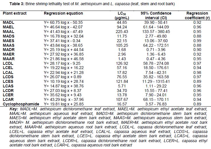
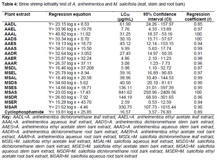
Findings from this study revealed that M. aethiopicum root bark extracts was the most toxic against brine shrimp larvae with narrow LC50 range of 1.43 – 2.96 µg/mL as shown in . L. capassa aqueous stem bark (LCAS) extract exhibited cytotoxicity activity against brine shrimp with LC50 value of 5.71 µg/mL followed by ethyl acetate root bark (LCER) and aqueous leaf (LCAL) extract with LC50 values of 13.78 µg/mL and 17.86 µg/mL respectively as shown in . The A. anthelmentica extracts exhibited LC50 values below 100 µg/mL () against brine shrimp and therefore considered as toxic. However, the A. anthelmentica aqueous root bark (AAAR) extract exhibited higher cytotoxicity activity with LC50 value of 3.08 µg/mL followed by ethyl acetate root bark (AAER) extract which had LC50 value of 4.86 µg/mL. The A. anthelmentica ethyl acetate leaf (AAEL) and aqueous stem bark (AAAS) extracts displayed LC50 values of 7.76 µg/mL and 9.99 µg/mL respectively. With regard to M. salicifolia, the ethyl acetate root bark (MSER) extract were active against brine shrimp larvae with LC50 value of 2.59 µg/mL followed by dichloromethane leaf (MSDL) extract which exhibited LC50 value of 5.98 µg/mL as shown in . In addition, this study revealed that there was a significant difference (p < 0.05) in the concentration of some extracts tested as shown in and 6. Furthermore, the degree of lethality increased with increase in the concentration for all plants tested and the standard control.
Discussion
Validation of ethnomedical information of plants commonly used by ethnic groups has been a strategy in the discovery of novel bioactive secondary metabolites [13]. However, biological screening of plants for a targeted biological properties has also been regarded an optional strategy towards unveiling medicinal potential of plants. Despite these efforts, there are still some plants that have never been screened for biological potentials. That is why M. aethiopicum, L. capassa, A. anthelmentica and M. salicifolia growing in Tanzania were evaluated for antifungal and cytotoxicity activities. Leaves of L. capassa were reported by [14] as a remedy for skin infections. Since fungus is the main cause of skin infections in humans, it is therefore evident that L. capassa leaves extract is a potential antifungal herbal product. The MIC value of 0.78 mg/mL recorded by L. capassa aqueous leaf extract (LCAL) against C. albicans and C. neoformans provide evidence that polar compounds in the leaves extract of this plant is a potent antifungal agent. Furthermore, the high cytotoxicity (LC50 17.86 µg/mL) supports its use as topical antifungal agent. According to [15], plant extracts that are recommended in the drug discovery initiatives are those with MIC value of less than 1 mg/mL, and thus plant extracts that exhibited MIC value of 0.78 mg/mL in this study are potential source of drug templates. However, [15] further elaborated that extracts with low antimicrobial activity should also be reported as they can be incorporated with other extracts to improve it biological importance.
A. anthelmentica dichloromethane leaf (AADL) and ethyl acetate (AAEL) extracts had selectivity against C. albicans with MIC value of 3.13 mg/mL. Conversely, the aqueous leaf (AAAL) extract had selectivity against C. neoformans with same MIC value of 3.13 mg/mL. Earlier phytochemical investigations on the leaves of A. anthelmentica reported the presence of phenolics and terpenes [16]. Since terpenes have been reported to possess antifungal properties [17], it is therefore postulated that the antifungal properties exhibited by A. anthelmentica might be due to the presence of terpenes. Findings from this study are in line with the previous study conducted by [18] which reported that leaves of A. anthelmentica growing in Kenya exhibited high antifungal activity as compared with other parts of the plant. Another study conducted by [19] on antifungal activities of A. anthelmentica by disc diffusion method in the eastern part of Tanzania reported that the extract was inactive against C. albicans with an average inhibition zone of 15 mm.
Antifungal investigation of M. salicifolia revealed that, leaf extracts selectively inhibited the growth of C. neoformans with MIC value of 3.13 mg/mL. This suggests that leaves of this plant are potential source of drug leads for the treatment of fungal infections caused by C. neoformans. According to Godfrey et al [20], utilization of leaves is highly recommended for sustainability of plants as the use of roots and stems increases risk of plants extinction. These results are in agreement with the traditional use of M. salicifolia leaves by Pare people in the Northern Tanzania for treatment of skin diseases [21]. Regarding M. aethiopicum, none of the extract exhibited antifungal activity with MIC value below 3.13 mg/mL and therefore suggest that the plant does not contain active ingredients against selected fungal strains.
In the cytotoxicity assay, brine shrimp lethality test (BST) was used to predict the potential cytotoxicity effect of extracts from M. aethiopicum, L. capassa, A. anthelmintica and M. salicifolia. According to [22], the BST is the rapid, inexpensive and simple bioassay for testing plant extracts bioactivity which in most cases correlates with cytotoxic and antitumor properties. Additionally, [12] demonstrated that plant extracts with LC50 value greater than 100 µg/mL are considered as non-toxic while LC50 value less than 100 µg/mL as toxic. Findings from this study revealed that M. aethiopicum root bark extracts were the most toxic against brine shrimp larvae which is evidenced by LC50 range of 1.43 – 2.96 µg/mL. The highest susceptibility shown by brine shrimp larvae towards root bark extracts of M. aethiopicum suggests that the root bark is the potential antitumor agents. The L. capassa stem bark and leaf extracts demonstrated cytotoxicity activity against brine shrimp with LC50 value range of 5.71 – 17.86 µg/mL. These results collaborate with the previous cytotoxicity investigation study of the same plant growing in Tabora region, Tanzania which gave LC50 value of 17.8 µg/mL on brine shrimp [23]. Bark and leaves of this plant are therefore a potential anticancer herbal product. Interestingly, cytotoxicity results of L. capassa aqueous leaf (LCAL) extracts are consistent with the results of a standard anticancer drug cyclophosphamide which demonstrated LC50 value of 16.57 µg/mL.
In this study, the cytotoxicity results displayed by A. anthelmentica are in agreement with previous study conducted by [24] where LC50 value below 100 µg/mL was reported for the root bark extract of the same plant species. The Myrica salicifolia ethyl acetate root bark (MSER) and dichloromethane leaf (MSDL) extracts demonstrated a potential source of antitumor agent as evidenced by LC50 values of 2.59 and 5.98 µg/mL respectively. The sensitivity shown by brine shrimp against ethyl acetate root bark and dichloromethane leaf extracts provide a circumstantial evidence that secondary metabolites in the root barks and leaves of M. salicifolia might be a good source of anticancer compounds. According to Moshi et al [23] a plant extract with LC50 value below 20 µg/mL have a likelihood of yielding anticancer compounds. Previous cytotoxicity investigation of M. salicifolia extracts in Kenya proven that aqueous extracts had high cytotoxicity against brine shrimp larvae [25]. These findings however, collaborate with the present findings, despite the geographical separation of the plant.
Conclusion
The extracts of L. capassa, A. anthelmintica and M. salicifolia exhibit vaying degrees of antifungal activities against C. albicans and C. neoformans whereas extract of M. aethiopicum displayed the least antifungal activity compared with the rest of the plant extracts tested. A majority of the extracts confirmed to be toxic and thus possess anticancer activity.
Declarations
Acknowledgement
References
Archives


News Updates